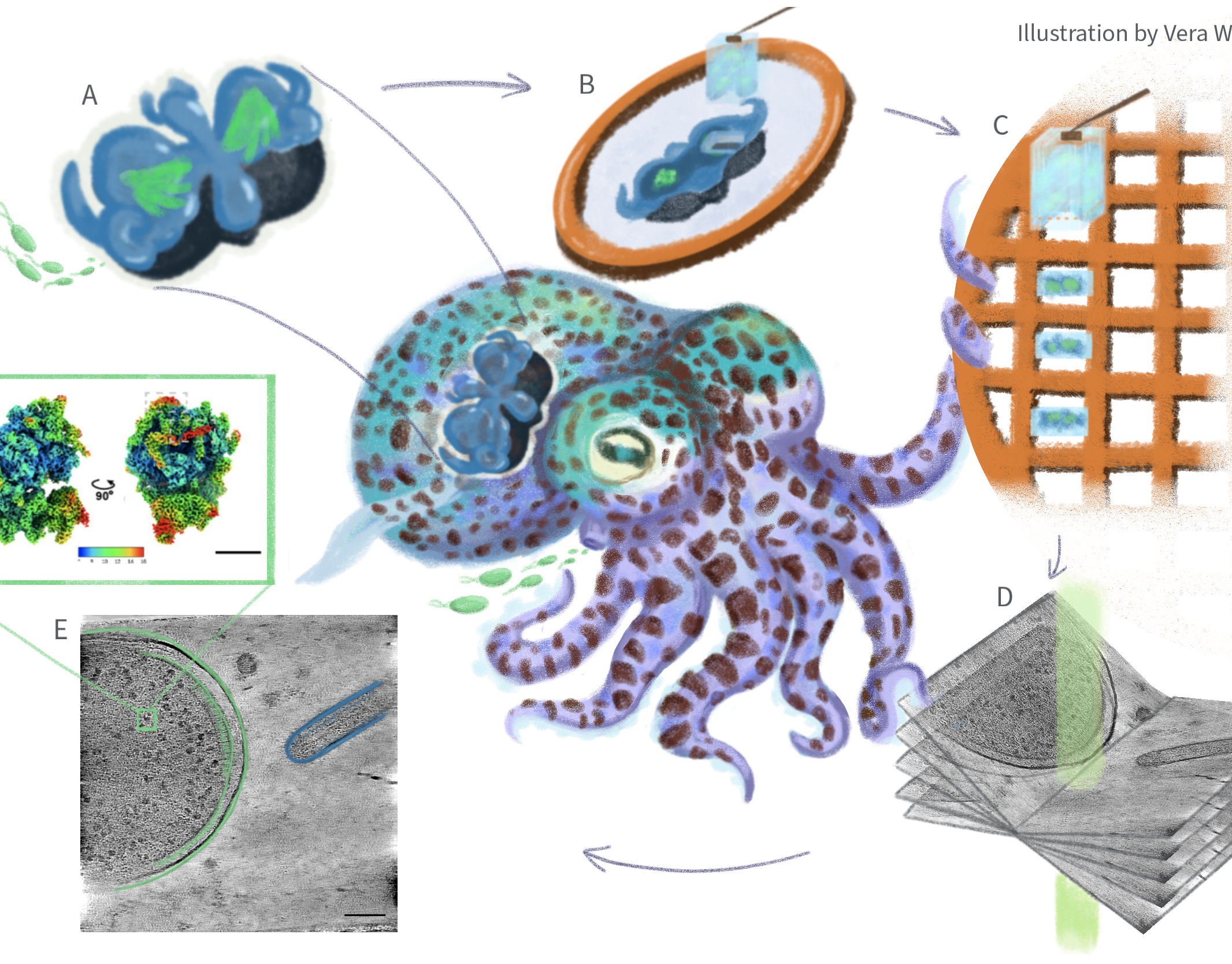
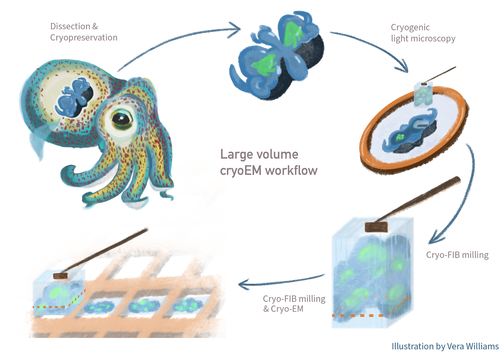
This header illustration was created for a publication on the large-volume Cryo-EM workflow of the squid light organ by Katrina Gundlach. It demonstrates how she gets her samples ready for imaging in the high resolution microscopes, and this is not an easy task! Although the samples seem small to us, they are still huge compared to how much we "zoom-in" under the microscopes. As such, multiple techniques are applied to make sure she gets the correct into view.
This work is part of the publication "Nanoscale imaging of native symbiotic animal tissue using a multimodal large volume imaging pipeline for cryo-electron tomography" and is currently in preprint.
Except where otherwise noted this work is protected under https://creativecommons.org/licenses/by-nc-nd/4.0/
References
Nanoscale imaging of native symbiotic animal tissue using a multimodal large volume imaging pipeline for cryo-electron tomography
Katrina A. Gundlach, Oda H. Schiøtz, Mark Ladinsky, Colin Raimann, Michael Rheinberger, Florian Beck, Barış Gündüz, Rob Langelaan, Martin Rücklin, Ronald W.A. Limpens, Edward G. Ruby, Margaret McFall-Ngai, Jürgen M. Plitzko, Ariane Briegel
bioRxiv 2025.11.30.691379; doi: https://doi.org/10.1101/2025.11.30.691379